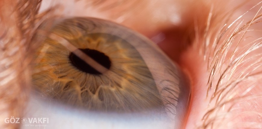
What is Retina? How is the inspection done?
The layer of the eye that detects light is called the retina. It completely encircles the eye's inner wall. It is made up of brain-derived vessels and nerves.
After measuring the degree of vision and passing the microscopic examination, drops are dripped to enlarge the pupil for the retinal examination.
Depending on the disease and the individual, the rate of pupil growth varies. Prior to the exam, this time period must be waited through.
The retina and intraocular transparent media (lens and vitreous) are then thoroughly examined with the aid of various tools.
What Tests Are Conducted for Retinal Patients' Diagnosis?
Biomicroscopic and indirect retinal examination.
Optical Coherence Tomography ( OCT )
Color Fundus Photo
Fundus Foleresin Angiography (FFA)
Green Angiography of Indosia (ICGA)
An Amsler card is given to those who are at risk of macular degeneration, and they are asked to look at it periodically. They are asked to come in right away for an examination if they notice their vision is crooked.
What signs do patients with retinal disease exhibit?
In retinal diseases;
Reduced vision,
Blurry or distorted vision,
Partial or sporadic vision
Floats in black,
Shadowing, flashing,
Low vision or even no vision complaints can happen in both light and darkness.
What are the signs and symptoms of retinal detachment?
There have been complaints about shadowing, flying, and flashing light in the eye. The two most significant risk factors are extreme myopia and eye trauma.
Laser treatment is used to stop the tear from progressing if it is discovered early on. The retinal layer is torn away from its position if there is a delay or negligence in diagnosis and treatment. Surgery is the only available treatment for this condition, which we will refer to as retinal detachment.
Which vision problems are associated with diabetes?
Numerous eye conditions are brought on by the systemic disease diabetes. It speeds up the development of cataracts and causes changes to eyeglasses, eye muscle paralysis, and double vision.
The destruction of the retina (nerve) layer in the eye is the most significant. Hemorrhages in the retinal layer, vascular occlusions, new vessel formations, and retinal edema are all signs of this condition, known as diabetic retinopathy.
How frequently should diabetics be examined? How is its treatment?
Patients with diabetes who have not yet begun to damage their eyeballs are advised to have an annual eye exam. Every six months, patients who have developed diabetic retinopathy should be checked on.
To assess the severity of diabetic retinopathy, fundus fluorescein angiography (FFA) and/or optical coherence topography (OCT) films may be taken if the damage has advanced. Laser treatment is necessary if there are areas of the retina layer with impaired nutrition, if new vessels are forming, or if there are leaking vessels. Medication is injected into the eye when the leakage around the yellow spot is frequent and cystic.
Bleeding that fills the eye occurs in untreated cases. In these patients, vitreoretinal surgery is used to treat hemorrhages. Shrinkages, membrane separations, and laser completion all take place during the procedure.
Treatments for diabetic retinopathy aim to preserve the degree of vision at the time of treatment initiation. Visual improvement is uncommon. Blindness occurs if treatment is forgone.
What kind of a disease is Yellow Spot Disease(Macular Degeneration)?
Over the age of 70, age-related macular degeneration affects about one-third of the population. It makes it difficult to read closely and especially with central vision. Vision deterioration and spotting develop over time in the center.
In later stages, vision in patients with initially distorted or crooked vision may go from being able to see a few meters to counting fingers.
Vision can be impaired to the point where it is considered legally blind, even though it is not completely blind. Patients lose their ability to focus on the object they are looking at, which prevents them from seeing a person's face in front of them but allows them to see their arms and legs. Such patients hardly have the ability to travel alone. Even though they are capable of working independently at home, they occasionally need assistance. They cannot read or write because they are blind.
There are two types of this ailment: the dry type and the wet type. In 80–85% of these patients, the dry type is present, and vision gradually deteriorates over years. As a course of treatment for these patients, protective and antioxidant pills are advised. In the case of the wet type, bleeding and edema quickly impair vision by developing at the visual point. Patients with this wet form of yellow spot degeneration are the ones we are treating right now.
Is Yellow Spot Disease Treatable?
Ten years ago, we were unable to offer these patients much assistance with their treatment. After the 2000s, numerous treatment modalities emerged. These advancements, which began with photodynamic therapy (laser application using intravenous drug administration to the patient), have persisted to the present day with the application of various substances injected into the eye. Aside from the medication used around the world that recently arrived in Turkey and had the opportunity to be applied to patients, the possibility of blinding the problem from this illness will finally be reduced to a minimum with the discovery of more effective drugs and methods in the near future as well as numerous researches that are still ongoing quickly.
Hereditary and metabolic disorders are believed to be the two most significant causes of this illness, despite the fact that its exact origins are unknown. In the future, individuals who will have this condition will be able to be treated with gene therapy or other methods of treatment before the disease manifests itself thanks to tests that will be performed at a young age.
Who Needs Regular Eye and Retinal Examinations?
Those who have diabetes (diabetes) themselves or in their family,
Patients with hypertension,
Elevated cholesterol,
Those suffering from heart disease.
Those who either personally or through their family have the yellow spot disease.
Those who have a history of inherited or congenital vision problems.
What is Retinopathy of Prematurity (ROP)?
ROP is a condition that affects low birth weight, premature babies, and can cause blindness.
What Are the ROP Risk Factors?
All infants delivered weighing less than 1300 g or before 30 weeks.
Babies receiving oxygen therapy in the intensive care unit who weigh less than 1500 g or were born at less than 32 weeks.
Babies at risk for ROP include those born prematurely with recurrent apnea, sepsis, bronchopulmonary dysplasia, respiratory distress syndrome, or ductus arteriosus patency, as well as babies whose mothers have preeclampsia and diabetes.
What is the procedure for ROP inspection and follow-up?
The ROP examination involves enlarging infants' pupils while giving them topical anesthesia so that the retina—the eye's nerve layer—can be seen. Premature infants should undergo their first ROP examination 4–6 weeks after delivery. The examination is then repeated every one to two weeks until the baby is full-term, depending on the presence and severity of the disease (time to be born).
What ROP treatment options are there?
Regular follow-up is the most crucial step in the treatment of ROP. According to statistics, 80% of patients who develop ROP spontaneously regress, and only 8% of infants who are monitored need treatment. However, the most crucial factor in preventing blindness in infants with ROP is early disease detection, or what we refer to as threshold disease, and urgent laser treatment (within 3 days). When the disease cannot be treated with laser therapy and cannot be detected in time, a vitrectomy is performed.
Photodynamic Treatment (PDT)
Some patients with macular degeneration (macular degeneration), chronic central serous chorioretinopathy, and polypiodal choroidal vasculopathic cases receive photodynamic therapy.
After administering a photosensitive chemical substance intravenously, the idea behind photodynamic therapy is to destroy the target tissue by stimulating it with low-energy light (cold laser), while making sure to occlude the vascular component in the target tissue without harming the surrounding tissues.
Repeated photodynamic therapy is an option if necessary. If the doctor decides it is appropriate, it is applied to both eyes simultaneously if there is damage to both.
Patients who receive treatment are asked not to expose themselves to harsh light for 48 hours.
You can contact our hospital with any type of eye health concern, and we'll provide you with thorough information about your eye issues as well as the steps involved in getting them treated.
You can contact our hospital with any type of eye health concern to receive detailed information about your eye issues as well as to learn the steps involved in treatment.


