Refractive Surgery
Refractive (Focusing) Defects and Treatment Methods
The eye is an organ basically similar to a camera. The rays coming from the external environment pass through the refractive surfaces of the eye such as cornea (the outermost transparent layer), lens and focus in the macula (yellow spot) region responsible for sharp vision in the network layer. The focusing of the rays in a different place is called refractive error. Refractive errors can be analysed in 3 different groups as myopia, hypermetropia and astigmatism.
Apart from these, the decrease in near vision caused by the decrease in the ability of the eye lens to focus the near over the age of 40 is called presbyopia. This condition is not an eye disease, but a natural part of the aging process.

MYOPIA: Myopia, defined as the inability to see far away clearly, is the result of the rays coming into the eye focusing in front of the retina. It most commonly develops due to the anterior-posterior axis of the eye being longer than normal. It is a structural feature and genetic transmission is common. It usually starts at school age and increases as the growth process continues. The development of cataracts in advanced age can also change the refractive power of the lens and cause myopia.

HYPEROPIA: In general, nearsightedness is not good. It occurs as a result of the focusing of the rays coming into the eye behind the mesh layer. In contrast to myopia, here the eye structure is smaller than normal. Structural and hereditary characteristics are the most common cause of hyperopia. Untreated hypermetropia increases the risk of lazy eye in children. Therefore, all children before school age should have an eye examination.

ASTIGMATISM: It is caused by the different refractive power of the cornea in different meridians. Astigmatism is an eye condition that causes blurred vision at any distance. It may be structural or may occur as a result of degenerative diseases, infections and traumas that can cause changes in the corneal layer.
PRESBYOPIA: It is a nearsightedness problem seen over 40 years of age. The lens inside our eyes has a structure that can change shape. Thanks to this special ability, the intraocular lens increases its refractivity by swelling when objects are close and we can see nearby objects clearly. After the age of 40, the eye begins to lose this ability gradually and the near vision difficulty called presbyopia begins.
Why Does Presbyopia Occur?
It is a nearsightedness problem seen over 40 years of age. The lens inside our eyes has a structure that can change shape. Thanks to this special ability, the intraocular lens increases its refractivity by swelling when objects are close and we can see nearby objects clearly. After the age of 40, the eye begins to lose this ability gradually and the near vision difficulty called presbyopia begins.
Prepared by the Editorial Board of Eye Foundation Hospitals.
EXCIMER LASER
Refractive errors can now be treated permanently with today's technology. This treatment, which has been safely applied all over the world for more than 30 years and has eliminated the dependence of millions of people on glasses or contact lenses, is performed with a device called Excimer laser. Excimer laser is a device that produces ultraviolet light at a wavelength of 193 nm using Ar F gas. The laser beam formed here eliminates the targeted tissue in the desired thickness and width, so that a permanent change occurs in the cornea layer located at the outermost part of the eye and myopia, hyperopia or astigmatism is treated. Femtolasik is the most commonly used method for this purpose.
LASIK
It is the most commonly used method in the treatment of focusing defects. Patients who are over 18 years of age and whose spectacle number has not changed in the last 1 year are candidates for this treatment. In order for Lasik to be applied, the number of glasses and the structure of the eye must be appropriate. For this purpose, all candidate patients undergo a detailed eye examination as well as a series of examinations showing the properties of the corneal layer. Myopia up to 12.0 degrees, hyperopia up to 6 degrees and astigmatism can be treated with LASIK. It is important to stop using contact lenses before LASIK. Patients using soft lenses should not wear lenses for at least one week, and those using hard lenses should not wear lenses for at least three weeks.
To whom lasik is not applied:
- - Those with corneal thickness below a certain limit
- - Those with severe dry eye
- - Diabetics and those with rheumatic diseases
- - Pregnant and lactating women
- - Those with conditions such as cataract, glaucoma, infection in the eye
Application
In the LASIK method, the cornea layer, which is located at the outermost part of the eye, is numbed with 1-2 drops of anaesthetic drops. Thus, the patient does not feel any pain during the intervention.
A speculum is placed on the lids to prevent involuntary lid movements. Then, a smooth surface is provided in the eye with a vacuum ring and an incision of approximately 0.12 mm is made in the corneal layer by means of a tool called microkeratome. The flap is removed and Excimer laser is applied to the underlying corneal tissue. Finally, the flap is placed in the same area and the operation is terminated. All these procedures are performed in an average of 5 minutes for both eyes and no pain is felt during this process.
The eyes are not closed after LASIK, it is recommended to rest the eyes as much as possible on the same day. Eye drops are used for an average of 1 week. During this period, it is not recommended to swim in the sea or pool, apply eye make-up and rub the eyes vigorously.
Advantages of LASIK treatment:
The most important advantage of LASIK treatment is the comfort it provides to the patient. In patients with appropriate eye structure, LASIK method can effectively correct even high spectacle degrees. Recovery is extremely fast and the patient can have a good quality vision within hours after the treatment. Pain, watering and stinging problems disappear within 12 hours at the latest and 1 week of medication is sufficient after the treatment.
Complications:
LASIK treatment is a treatment that takes place in the outermost layer of the eye. Therefore, it is not possible to damage the structures inside the eye. In addition, very rarely, some problems may occur during the removal of the flap from the cornea, as well as problems such as infection, inflammation, allergy and inadequate correction of the degree after treatment. These problems can be minimised with the experience of the physician performing LASIK treatment and the quality of the laser device used. The quality of the Excimer laser device, especially FDA approval, should be questioned before having Lasik.
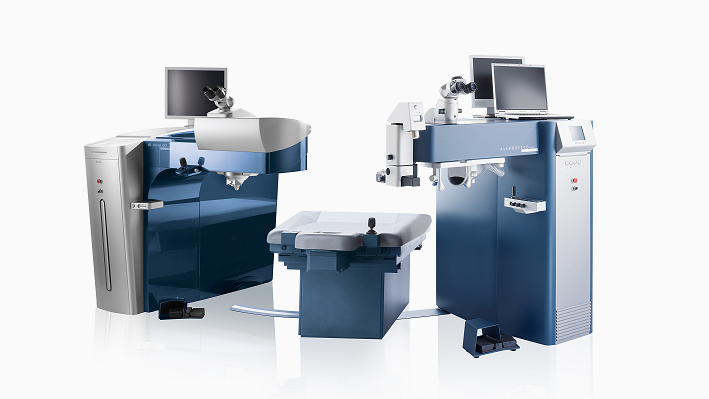
Alcon ALLEGRETTO WAVELIGHT EX 500, which is used in our hospital, is one of the fastest laser systems in the world with a speed of 500 Hz. It can treat 1 dioptre eye defect in less than 1.5 seconds. Thanks to the eyetracker (eye tracking system) system at 1050 Hz speed, laser treatment is extremely safe.
FEMTOLASIK
The most important step in LASIK treatment is the creation of a flap called flap from the upper part of the cornea. In recent years, femtosecond laser technology has been used to create the flap. Femtosanelaser is a device that can create incisions in the desired depth, width and shape in the tissue. The FS 200 Femtosanelaser is the fastest and safest technology developed for this purpose.
Advantages
- - This device has virtually eliminated the complications associated with previously used microkeratome systems.
- - With femtosecond laser, special flaps can be created for the person's refractive error.
- - Especially in the treatment of hyperopia and astigmatism, the results are more successful.
- - Corneal biomechanics are better preserved in femtosecond laser flaps.
- - Since it can lift the flap in the desired thickness, Lasik can be applied especially in patients with corneal thickness at limit values.
- - Since the phlebar surface is smoother, less aberration (light deviation) develops after laser treatment.
- - Femtosecond fluorescence lamps have a lower risk of developing dry eye, especially in elderly patients.
- - With femtosecond laser systems, besides flap formation, corneal implants can be inserted, keratoplasty (corneal transplantation) and astigmatic keratotomy procedures can be performed.

SURFACE TREATMENTS : (PRK-LASEK-NO TOUCH)
In the treatment of refractive errors with excimer laser, LASIK (the method applied by removing the flap from the cornea) is applied as the standard treatment all over the world. However, the corneal structure of each patient may not be suitable for this treatment. In such cases, surface treatments should be preferred. The biggest disadvantage of these treatments, which have the same long-term results as LASIK when the appropriate method is selected, is that the recovery time is long and corneal haze called haze may occur in high corrections. Surface treatments, which have been applied less frequently in recent years with the widespread use of femtolasic treatment, are still a highly effective and safe treatment alternative, especially in the correction of low numbers.
WAVEFRONT TREATMENT
In laser treatment, a laser correction is made only on the basis of the patient's degree of spectacle. In wavefront treatment, the wavefront device measures the light scattering in the cornea, lens and nerve layer in detail and organises a treatment scheme according to this measurement. The treatment scheme calculated in the Wavefront device is transferred to the laser device and thus personalised laser treatment is performed. Wavefront treatment is a method that should be preferred in patients with high sequential aberration rate in the measurements made before Excimerlaser treatment.
LENTICLE REMOVAL THROUGH A SMALL INCISION:
It is a method of creating a corneal stromalenticule in the corneal stroma with femtosecond laser and removing it mechanically. It can be used in the treatment of myopia and myopic astigmatism. Compared to LASIK, the refractive results are similar, postoperative dry eye is less common because the incision is smaller. The disadvantages are that the success rate decreases in the presence of high astigmatism, and it cannot treat refractive errors such as hyperopia, hyperopia astigmatism and mixt astigmatism.
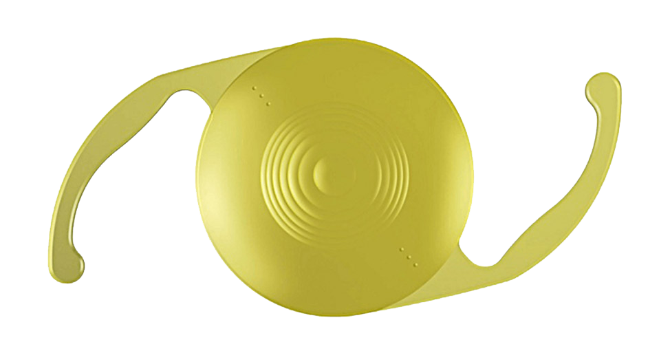
PHAKIC LENS
It is a preferred method for people between the ages of 20-45 whose corneal structure or degree of refractive error is not suitable for Excimerlaser treatment.
There are 2 different types of phakic lenses that can be fitted to the anterior or posterior chamber.
- - Fixated on the iris
- - Settled in Sulcusa
In order for these lenses to be fitted, personal characteristics such as endothelial number and anterior chamber depth must be appropriate.
REFRACTIVE LENS REPLACEMENT
Over 45 years of age, it is a method used in the treatment of myopia, hyperopia, astigmatism and presbyopia (near focusing difficulty due to old age). Thanks to the Toric (correcting astigmatism) and Multifocal (providing both near and far vision) lens technologies that have developed especially in recent years, we are able to offer our patients a life without glasses.

Refractive Surgery Frequently Asked Questions
Does pain occur during and after laser treatment?
Laser treatments are performed with the help of eye drops with anaesthetic properties. No pain is felt during the operation. Since the procedure takes place in a very short time such as 5 minutes per eye, the operation is over before most of our patients realise what is happening.
Postoperative discomfort (stinging, burning, watering, blurred vision) in lasik treatment lasts approximately 5-6 hours. In other words, patients can easily fulfil their daily activities when they wake up one day later.
Things to consider before laser treatment?
Patients who wear hard contact lenses should stop wearing them 3 weeks in advance, and those who wear soft contact lenses should stop wearing them 1 week in advance. It is recommended to wear comfortable clothes, no make-up and even no perfume on the day of surgery.
Is laser treatment safe?
Excimer laser treatment has been applied all over the world for about 25 years. In this process, we have not encountered serious complications that would require us to stop this treatment. Especially with the help of the recently developed examination devices, the results are very satisfactory if the appropriate patient selection is made and the right treatment is applied at the right time.
What to do after laser treatment
Immediately after the treatment, 2 eye drops containing antibiotic and cortisone are given to our patients and they are asked to use them every hour. After one day, the doses are reduced and these drops are used for one week.
For 1 week, it is important not to enter the sea, pool and not to rub the eye.
Can a more perfect treatment emerge in this period of continuous technological development?
This question has always confused me as an ophthalmologist and caused me to wait for Excimer laser. However, the results we have obtained at the point we have reached now are so satisfactory that I thought that there was no end to waiting and that I had to catch the technology from somewhere and I had this treatment 8 years ago. I am extremely pleased with the decision I have made now.
Is laser treatment an obstacle for future cataract surgery?
Before cataract operation, a measurement called biometry is performed to calculate how many intraocular lenses will be implanted. Since excimerlaser treatment permanently changes the corneal curvature, there were difficulties in intraocular lens calculations for a while. However, this problem has been overcome with new formulae developed in the last 4-5 years. Therefore, all eye surgeries, including cataract surgery, can be performed safely after Excimerlaser treatment.
Is there a difference between femtosecond Lasik and normal Lasik?
Femtosecond laser is a method that creates a flap (a piece of tissue removed from the upper part of the cornea), which is the most important stage of Excimer laser treatment. It can also be used in corneal ring applications and kerotoplasty (corneal transplantation) surgeries. This Nobel Prize-winning laser technology creates a very smooth incision in the cornea with infrared pulses in a quadrillionth of a second. Complications that can be seen in standard keratomas during flap formation almost do not occur in this system, so it is a safer method. There are publications reporting that refractive surgery using femtosanelaser gives better visual results compared to standard keratome treatment.
As a result, Excimer laser surgery is an aesthetic surgery. It is not a mandatory procedure. Therefore, when surgery is decided, it is necessary to perform this work with the safest possible method. Excimer laser treatment with femtosecond laser flaps is the most reliable method for today.
Is it possible to regain the number after laser treatment?
It is not possible for the entire corrected number to come back, but 5 out of 100 patients may have numbers that require the use of glasses. In this case, which is usually encountered in high number corrections, it is possible to re-laser from the same area if the corneal structure allows.
Is it possible to cure astigmatism?
Since the excimerlaser treatment started, it is possible to treat astigmatism up to number 6.
Will the treatment fail if I move my eye during laser?
Thanks to the eyetracker, i.e. eye tracking systems in Excimerlaser devices, the slightest movements of the eye are detected and the treatment is directed to the correct area. If there is a movement that the device cannot follow, the treatment is stopped. Therefore, it is not possible to have a bad result due to the patient's non-compliance.
Is the no touch treatment a newer and more successful treatment?
No touch or transepithelial PRK is a treatment that has been available since the beginning of Excimerlaser applications and is included in the surface ablation group. What is done here is to somehow remove the epithelial layer located at the outermost part of the cornea and apply Excimerlaser directly. The epithelium can be removed by scraping, with the help of alcohol or with special devices developed for this purpose, or it can be removed with Excimerlaser. The results of the treatments in the surface ablation group are the same in terms of predictability, safety, stability and there is no difference in terms of complications that may occur in the studies. Surface ablations are currently preferred only in patients with low corneal thickness due to the long rehabilitation period and less predictable refractive results compared to LASIK, especially at high numbers.

